Western blotting has been around for more than 35 years and remains one of the most heavily utilized techniques in biological sciences. And lately, a lot has been written on how to make western blotting more reproducible. These include: monitoring the protein loading, optimizing transfer conditions, conducting antibody validation, replacing the autoradiographic film with the digital imaging, and automating the process, among others.
What’s missing here is the answer to one important question:
How do we know that our protein of interest is, in fact, present on the membrane?
Most of us scientists take for granted the fact that once transferred, the proteins are stably bound to the membrane and will not come off. To challenge this assumption, it is important to remember that proteins are likely adsorbed onto the membrane via the dipole and hydrophobic interactions. Therefore, given the differences in charge across the different proteins, it is plausible to assume the protein adsorption and retention would not be uniform.
To maximize the chances of the proteins being adsorbed onto popular membranes such as Polyvinylidene Fluoride (PVDF), pre-wetting the membranes in methanol prior to the transfer is a well-known step.
However, the correct post-transfer steps are barely emphasized to avoid protein signal loss on the final blot.
Given the time constraints, many researchers put the membrane directly into the blocking buffer, without drying and chemically fixing it. In those cases, the proteins that are not tightly bound to the membrane detach upon shaking, thus leading to the signal loss. Consequently, the tempting, yet only partially correct explanation for the signal loss, is that shaking removes the protein off the membrane (1).
A Western Blot Protein Signal Loss Prevention Strategy:
To retain the proteins on the PVDF membrane post-transfer, prior to the subsequent blocking, washing, and incubation with antibodies (2):
1. First, air-dry the membrane.
2. Then, place it into a small amount of 100% methanol to wet the membrane.
3. Then place it in distilled water to remove the methanol.
This largely overlooked, yet critical detail has been described as far back as in 1987 for the PVDF membrane (3). Subsequently, several articles have demonstrated that the post-transfer exposure of PVDF membranes to methanol, glutaraldehyde or paraformaldehyde was essential to detect several proteins (4-6).
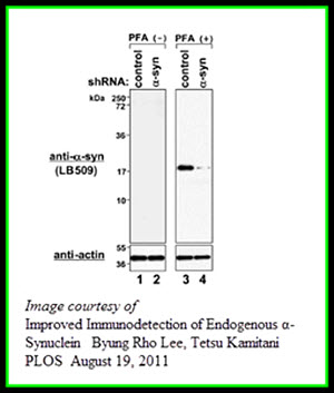
Although post-transfer treatments are not generally done for the other widely used nitrocellulose membranes, exposure to paraformaldehyde has shown to be beneficial in at least one study (5). The exact reasons for this phenomenon may vary.
However, it is safe to hypothesize that the combination of water removal by air drying and chemical fixation would lower the detachment of the proteins during shaking associated with the washes/antibody incubation. In this way, keeping the proteins attached to the membrane would be yet another essential part of making the results of your Western blotting experiments more reproducible.
Western blotting remains indispensable in measuring and detecting proteins in biological samples. The individual steps are quite simple, yet the devil is in the details.
If you don’t see the protein expression by Western blotting even after trying multiple antibodies and other protocol optimizations, don’t despair.
The reason could be as simple as the protein loss from the membrane. And, fortunately, in most cases, this is easy to remedy.
By spending as little as an extra five minutes of your time using our Western blot protein signal loss prevention strategy, in most cases, you will be able to to properly process the membrane post-transfer.
But remember: sometimes preventing protein signal loss from your Western blot depend upon the choice of membrane.
Should it be PVDF or Nitrocellulose? For Help in Choosing the Right Membrane for your Western blot, check out
Insane in the Membrane! PVDF vs. Nitrocellulose – Which One Comes Out on Top?
and
The Membrane Matters
References:
- Tomisawa, S., Abe, C., Kamiya, M., Kikukawa, T., Demura, M., Kawano, K., and Aizawa, T. (2013). A new approach to detect small peptides clearly and sensitively by western blotting using a vacuum-assisted detection method. Biophysics 9: 79-83.
- Ni, D., Xu, P., and Gallagher, S. 2016. Immunoblotting and immunodetection. Curr. Protoc. Mol. Biol. 114:10.8.1-10.8.37.
- Matsudaira, P. (1987). Sequence from picomole quantities of proteins electroblotted onto polyvinylidene difluoride membranes. J Biol Chem 262 (21): 10035-10038.
- Mizzen, C., Cartel, N., Yu, W., Fraser, P., McLachlan, D. (1996) Sensitive detection of metallothioneins-1, -2 and -3 in tissue homogenates by immunoblotting: a method for enhanced membrane transfer and retention. J. Biochem. Biophys. Methods 32: 77-83.
- Suzuki, Y., Takeda, Y., Ikuta, T. (2008) Immunoblotting conditions for human hemoglobin chains. Anal Biochem 378: 218–220.
- Lee, B., Kamitani, T. (2011) Improved immunodetection of endogenous a-synuclein. PLoS ONE 6(8): e23939.


